Faculty of Medicine and Health Sciences
Department of Circulation and medical imaging
 |
 |
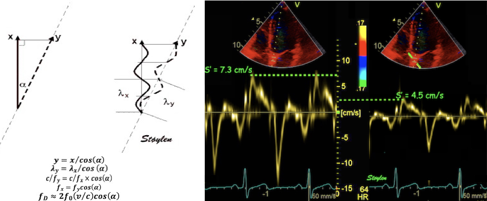
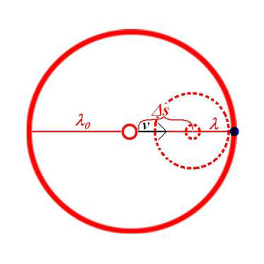
| The Doppler
effect for a moving source and a stationary
observer. In the time the original wave has moved
a wavelength |
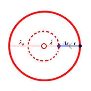
The Doppler effect for
a stationary wave source and a moving observer. In
the time the wave has moved the distance |