482f Low Frequency Sonophoresis: Ultrastructural Basis for Stratum Corneum Permeability Assessed Using Quantum-Dots
Here we report, using quantum dots (QDs) as a tracer and confocal microscopy and transmission electron microscopy (with OsO4 and RuO4 post-fixation) as visualization methods, the nature of LFS-induced permeation pathways in the skin. Confocal microscopy studies showed that LFS delivered QDs (20 nm diameter) across the stratum corneum, forming transport regions spanning about 40-80 microns in width and upto 60 microns deep. Transmission electron microscopy (TEM) revealed that QDs penetrated within the lipid regions of stratum corneum, the intercellular spaces at the stratum granulosum-stratum corneum interface, and the nucleated layers in the epidermis. TEM microgrpahs of control as well as ultrasound-treated skin showed the presence of voids in the intercellular lipid regions, referred to as lacunae. However, whereas the lacunae were few in number and more scattered in controls, their size and density was significantly increased in ultrasound-treated samples. Quantitative image analysis of electron-micrographs showed a significant increase in lacunar dimensions when 1% w/v SLS was added to the coupling medium. These ultrastructural studies show that LFS induces dilatation and higher connectivity of voids in the stratum corneum possibly leading to formation of a three-dimensional porous network which is capable of transporting QDs as well as macromolecules across the stratum corneum.
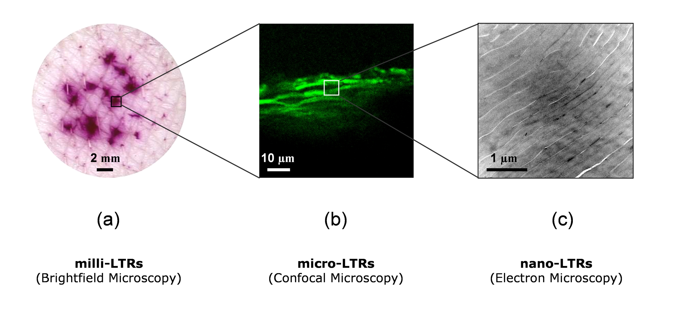
Figure Caption: Highly heterogeneous manifestation of low frequency ultrasound induced localized transport regions (LTRs) in the skin at different lengthscales. (a) Macroscopic brightfield scan of skin surface showing patches of milli-LTRs imbibed with a colorimetric dye, Sulforhodamine B. (b) Confocal-micrograph of micro-LTRs showing preferential penetration of quantum dots within milli-LTRs. (c) At a nano-scale, the transmission electron-micrograph shows the existence of nano-LTR pockets of QD penetration in the stratum corneum.