160g In Vitro Cytotoxicity Studies of Oxides Nanoparticles and Comparison to Asbestos
We have therefore evaluated a human mesothelioma and a rodent fibroblast cell line for in vitro cytotoxicity tests using industrially important nanoparticles. Their response in terms of metabolic activity and cell proliferation of cultures exposed to 0 to 30 ppm nanoparticles (µg g-1) was compared to the effects of non-toxic amorphous silica and toxic crocidolite asbestos. Solubility was found to strongly influence the cytotoxic response. The results further revealed a nanoparticle specific cytotoxic mechanism for uncoated iron oxide and partial detoxification or recovery after treatment with zirconia, ceria or titania. While in vitro experiments may never replace in vivo studies, the relatively simple cytotoxic tests provide a readily available pre-screening method.
References: T.J. Brunner, P. Wick, P. Manser, P. Spohn, R.N. Grass, L.K. Limbach, A. Bruinink, W.J. Stark, In vitro cytotoxicity of Oxide Nanoparticles: Comparison to Asbestos, Silica, and effects of particle solubility. Environmental Science & Technology (2006), published online, 2006, DOI: 10.1021/es052069i
L.K Limbach, Y. Li, R.N. Grass, T.J. Brunner, M.A. Hintermann, M. Muller, D. Gunther, W.J Stark, Oxide nanoparticle uptake in human lung fibroblasts: Effects of particle size, agglomeration, and diffusion at low concentrations. Environmental Science & Technology 39, 9370-9376 (2005).
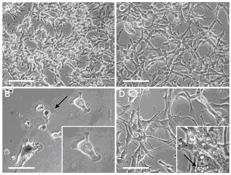
Figure 1. Morphological changes of MSTO cell cultures after 6 days incubation. A control cells without treatment; B 7.5 ppm crocidolite (see arrow for a single fiber), enlargement of a single cell which has increased in size, fibers surrounding the cell; C 7.5 ppm ZrO2, no visible changes in morphology; D 15 ppm ZrO2 resulted in the expression of small vesicles (probably apoptotic bodies, see arrow) in some cells; Scale bar 100 µm.
