477b Metal-Polymer Nanocomposites for Therapeutic and Imaging Applications
Research in the field of biomaterials and drug delivery is moving toward individually tailored intelligent therapeutic systems which are capable of responding to and correcting undesirable conditions, on a molecular level, within the body.Ā However, to date no one single system has been able to successfully combine the aspects of biocompatibility, intelligent response, imaging, and non-invasive external therapeutic control.Ā Therefore, in this paper we explored the development of a metal-polymer nanocomposite system capable of synergistically incorporating all of these properties together into one complete intelligent therapeutic system.
|
|
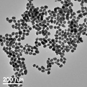
| Figure 1. TEM micrograph of gold nanoparticle after preparation (d ~ 40 nm). |
The intelligent therapeutic nanocomposite systems presented here are composed of a metal nanoparticle core (~40 nm diameter), surrounded by a temperature-responsive interpenetrating polymer network (IPN) layer (~150 nm thickness), which encapsulates desired molecules or compounds, and exhibits a sharp positive swelling transition, which is ideal for drug delivery applications.Ā The metal nanoparticle core of these systems can be tuned to absorb visible to near infrared (530 ¢ 1200 nm) wavelengths of light depending on either the core to shell thickness in metal nanoshells or the diameter in solid metal nanoparticles.Ā This absorbed light energy is then converted to heat and transmitted locally to the surrounding IPN layer, which triggers the swelling of this temperature-response polymer layer and hence the triggered release of any encapsulated agents.Ā These metal nanoparticles can also be irradiated with nanosecond pulsed laser light which when absorbed leads to the production of broadband ultrasound waves that can then be subsequently used for photoacoustic imaging of the nanocomposite systems within body.
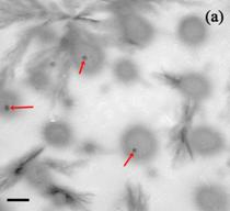
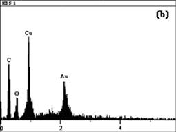
The size and relative shape of the prepared gold nanospheres was examined using transmission electron microscopy (TEM) and is illustrated in Figure 1.Ā These gold nanospheres were subsequently incorporated into IPN nanoparticles by in-situ emulsion polymerization.Ā After polymerization the successful entrapment of gold nanoparticles within IPN polymer particles was confirmed using TEM and Energy Dispersion Spectroscopy (EDS) on an individual metal-polymer nanocomposite particle as shown in Figures 2.Ā
|
|
|
| Figure 2. (a) TEM micrograph showing the presence of gold (small dark circles, indicated by red arrows) inside the lighter grey colored polymer particles (scale bar represents 500 nm).Ā (b) EDS spectroscopy of an individual nanocomposite particle showing a carbon and oxygen from the polymer and a gold peak from the encapsulated gold core (copper peak is from the mounting grid). | |
Therefore, these results clearly illustrate the successful combination of metal and polymer nanoparticles together into a single biocompatible intelligent therapeutic system, capable of achieving an intelligent response, imaging, and non-invasive external control.
This work was supported in part by a grant from the National Science Foundation, CTS-03-29317, and by a National Science Foundation Integrative Graduate Education and Research Traineeship (IGERT) Fellowship (to D.E.O.), DGE-03-33080.