338d Combined Effect of Brain-Derived Neurotrophic Factor and Ecm-Coated Substrates on Neurite Extension and Persistence from Retinal Explants
Neurotrophins are a class of proteins that influence neuron adhesion, survival, neurite outgrowth and neurite persistence. Recently, neurotrophins have been examined as therapeutic agents for the treatment of catastrophic nerve injury and neurodegenerative disease, and also to improve the tissue-device interface of implanted neural prostheses. However, it is unclear if sustained delivery of neurotrophins will be required to achieve maximum effect. Long-term delivery of neurotrophins would likely be difficult, presenting similar technological challenges to insulin delivery.
Neurotrophins operate primarily through the activation of tyrosine kinase receptors, with their binding initiating protein signaling pathways. However, these pathways can also be influenced by the presence of extracellular matrix factors (e.g., laminin). If neurite regrowth could be initiated by neurotrophin delivery and sustained through the use of extracellular matrix factors, it would be possible to construct devices that produce long-term regeneration in the nervous system.
We examined the interaction between neurotrophins and substrates commonly used to promote cell adhesion and neurite outgrowth in a central nervous system model. Specifically, we examined the effect of brain-derived neurotrophic factor (BDNF) and laminin, collagen, poly-L-lysine, and CDPGYIGSR peptide-gold coatings on neurite density and length using retinal explants. These results were compared to control surfaces consisting of tissue culture treated polyester membranes and gold-coated polyester membranes. After 7 days, collagen surfaces produced the largest increase in neurite outgrowth, whereas outgrowth on control surfaces was minimal.
Additionally, we examined the persistence of neurites on ECM-treated surfaces following 7 days of BDNF withdrawal. Unlike previous reports using PC12 cells, a significant decrease in neurite outgrowth was not evidenced. In fact, neurite extension and number continued to increase even after BDNF removal. This effect was most pronounced for explants on collagen and laminin coated substrates.
These results suggest that ECM factors could be used to construct composite materials that can direct and sustain neurite outgrowth over time. Although in vivo studies will be required to examine longer time frames (e.g., > 2 weeks), retinal explants represent a close approximation of in vivo conditions. Retinal explants contain neurons (e.g., photoreceptors, retinal ganglion and bipolar cells) and glia (e.g., Mueller cells) as well as connective tissue. Few studies have examined synergistic effects between cell adhesion molecules and neurotrophins. Our results support their use in biomaterials to promote sustained neurite extension.
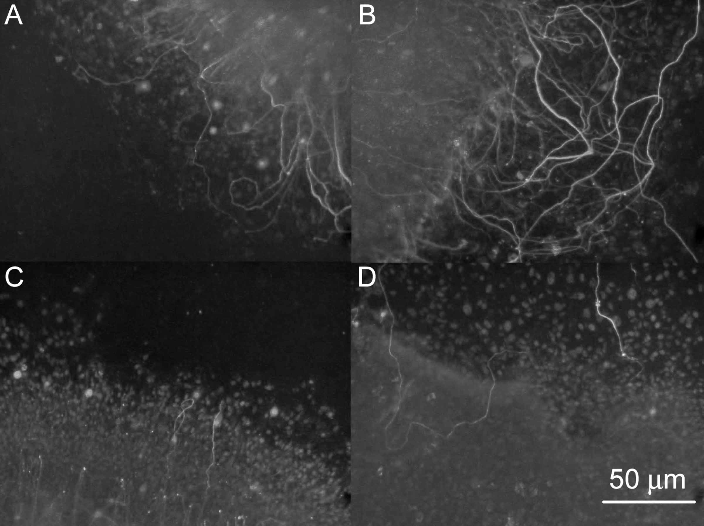
Figure 1: Neurite persistence following BDNF withdrawal on ECM-coated substrates. (A) Explants receiving BDNF and cultured on collagen-coated substrates for 7 days exhibited significant neurite extension and density when compared to (C) explants receiving BDNF and cultured on tissue-treated polyester control surfaces. On collagen (B), neurites persisted and increased in length and density even after 7 days of BDNF removal, whereas the density of neurites on control surfaces (D) appeared unchanged following BDNF removal.